Models for Routine Diagnostics
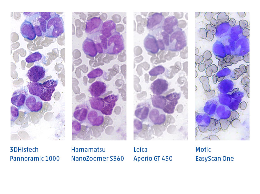
In the laboratory, the scanning process precedes digital diagnostics: After preparing the tissue samples, the slides are digitized in scanners before they are digitally diagnosed. But which scanner is the best? Which one is most suitable for routine diagnostics? Martin Weihrauch, M. D., hematooncologist and CEO of Smart In Media, has compared several models for the field of hematology:
 The desire to digitally microscope blood and bone marrow smears was the initial impetus for my intensive involvement with digital microscopy almost ten years ago. This is how the company Smart In Media, which I founded as a hematooncologist, became a specialist in this field. In the meantime, we have become the market leader in the field of digital pathology with our PathoZoom® products.
However, digital solutions have still not found their way into the routine diagnostics of us hematologists. This is mainly due to the lack of fast scanners for routine diagnostics in hematology. While our colleagues in pathology have long been able to enjoy good and fast devices with a resolution of 40x and scan times of about 1 minute per slide diagnostics, we hematologists still need to be patient to see our beloved slides digitally.
Even though the skilled pathologist will now probably roll his eyes at the claims of hematologists: Yes, we want a scan that makes our cytomorphology appear digital as if we were viewing the slides with an oil immersion objective at 60x, 80x or 100x in our microscope.
Manual scanning at the microscope in fantastic quality
This is already technically possible today, e.g. with our self-developed manual scanning solution PathoZoom® Scan in fantastic quality. Or also with the Olympus VS120 scanner. However, both options have the limitation that scanning dozens of slides per day would take forever. The Olympus VS120 needs about 30 minutes or longer for a scan of about 1×2 cm. A whole slide would take hours if necessary.
To illustrate how beautiful such a scan with our low-cost PathoZoom® Scan or with the Olympus VS120 can look, I have included cytomorphological slides for you to microscope.
Regarding manual scanning with PathoZoom® Scan, it is important to mention: for scanning a 40x oil with high aperture is sufficient, because the PathoZoom® camera has a higher resolution than the human retina, therefore you get almost the same result as if you were scanning at 100x oil.


