


The Big Microscope Scanner Test:
Speed, Quality, and Innovation in Digital Pathology

Martin Weihrauch, M.D.
Disclaimer: The reviews, opinions, and conclusions published in this test are based on our own experiences, research, and subjective assessments. They do not constitute a binding purchase recommendation and may vary depending on individual requirements and usage scenarios.
We assume no liability for the accuracy, completeness, or timeliness of the information provided. Changes to products, technical specifications, or prices may occur at any time. Please check official manufacturer information or independent sources before making a purchase decision.
The author is Chief Medical Officer at Smart In Media AG and is independent in his opinion regarding the microscope scanners presented. However, there is a conflict of interest regarding manual scanning, as PathoZoom® Scan & LiveView is a Smart In Media product.
Microscope scanners, also known as “Whole Slide Image Scanners” or “Digital Slide Scanners,” have been around for over 20 years.
Recently, there has been increasing momentum in the development and sale of digital slide scanners. Some say a “wave of digitization” is sweeping through microscopy, with pathology being the driving force. This is because no other field relies as heavily on microscopes as pathology. It is therefore only logical that digitization is now taking hold here as well—almost 20 years after the major wave of digitization in radiology, the other medical imaging-based discipline.
The Long Road to Digitization
There are three main reasons why radiologists have been working digitally for 20 years, while pathology is still in its early stages:
The main reason is that, apart from pathologists, few people understand what can be seen under the microscope in histology. In contrast, in radiology, not only radiologists need the images, but also surgeons and physicians from other specialties.
Another reason for the slow adoption of Digital Pathology is that, for a long time, there were no clear advantages of switching to digital imaging. Unlike radiology, where digital images completely replaced film, glass slides and staining are still required in pathology, and these materials are inexpensive.
The third reason for the slow digitization process is the high price of the necessary hardware—namely, the scanners.
However, the current shortage of pathologists is now driving the recruitment of specialists from remote locations, which is only feasible through digital remote work. Additionally, the increasing availability of artificial intelligence is prompting more institutions to transition to Digital Pathology.
For the first time, pathologists can diagnose cases faster (and more accurately) on a screen than under a microscope. With open-platform solutions like PathoZoom® Digital Lab by Smart In Media (the author is the company’s founder), switching between slides and cases happens with a mouse click—much faster than with a traditional microscope. Issues like poor posture, prolonged eye refocusing (70% of pathologists are over 50 and therefore presbyopic), mix-ups of glass slides, and misplaced requisition forms are becoming a thing of the past. Case reviews by senior pathologists are also significantly faster and smoother, as trainees can annotate key areas for their supervisors.
The key requirement for Digital Pathology is the microscope scanner, which consists of a microscope with one or more objectives, a digital camera, a motorized stage, and a loading mechanism for the slides.
The slide is automatically moved across the stage, position by position, while the digital camera captures images of each field of view. These individual images are then stitched together to form a single large image.
If a user scans a 2 x 1 cm area at 40x magnification, for example, the resulting image might be 80,000 x 40,000 pixels (3.2 gigapixels, or 300 times more detail than a 10 MP smartphone photo). These files typically range between 1-2 GB, meaning that with 300 slides per pathologist per day, massive amounts of data accumulate.
Currently, all high-throughput routine scanners remain in the higher price segment. Automatic scanners that can capture only one slide at a time (and are therefore unsuitable for routine workflows) typically start at $22,000 – $55,000 USD. For a high-throughput scanner from manufacturers like 3DHistech, Hamamatsu, or Leica, deep pockets are required—prices typically range from $110,000 – $270,000 USD (taxes not included).
Digitization Doesn’t Have to be Expensive
For those who want to wait a little longer or are not yet ready for full-scale digitization, there is an affordable entry point: manual scanning. Some products allow users to upgrade their existing microscope with a camera and computer software. The area of the slide to be scanned is moved manually under the microscope, creating a digital ‘Whole Slide Image’ (WSI).
The most affordable manual scanning solutions are available for under $16,000 USD. They are not suitable for digitizing dozens of slides per day, but they can cost-effectively and with excellent quality digitize individual slides, such as in a frozen section scenario.
Which Scanner for Which Situation?
First, it’s worth mentioning that for some applications, purchasing a scanner may not be necessary. This applies to teaching, where an institution wants to digitize a slide collection (e.g., 150 slides) and use it for years. If new slides are only added occasionally, it makes more sense to use a scanning service, which is available starting at $5–$25 USD per slide with excellent quality.
If the goal is to support remote consultation or a frozen section station for quick diagnostics, a full scanning system may not be required. In many cases, a more cost-effective ‘live viewing’ (streaming) system—a camera attached to an existing microscope combined with streaming software—can meet the need. Often, live viewing and manual scanning solutions are more flexible and faster than an automated scanner, as manual scanning at 4x or 10x magnification is often quicker than an automated scanner, which typically only has 20x or 40x objectives—taking longer than needed for rapid diagnostics.
For all other cases—where slides need to be scanned daily and analyzed asynchronously—an automated scanner is the best choice.
Single or Multiple Slides?
Microscope scanners are divided into ‘single loader’ and ‘multi loader’ models, but the real distinction should be between scanning fewer than 10 slides versus 10 or more slides at a time.
A scanner that can only load 1 to 10 slides at a time is not practical for pathologists handling 50 cases and 300 slides per day, as the loading and unloading process requires too much time and manual effort from lab technicians.
For high-volume workloads, a scanner with a large loading capacity (20+ slides) is necessary.
Speed Matters
Anyone who has observed a pathology lab at work might be reminded of the speed of a supermarket checkout. Pathologists can deliver a normal gastric biopsy diagnosis within 30 – 60 seconds, from microscopy to report generation (often using macro keys). Scanner and software developers should prioritize maintaining this speed rather than slowing it down.
In fact, speed, rather than image quality, has been the biggest bottleneck for microscope scanners so far. The good news: Advances in faster and more precise motorized stages and intelligent software algorithms are improving scanning speed, making Digital Pathology viable for routine use today.
Open Architecture vs. Locked-In Systems
Pathology labs already operate with a “mixed ecosystem” of scanners from different manufacturers and software from multiple vendors. To avoid compatibility issues between the Laboratory Information System (LIS), Digital Microscopy Software, and scanner hardware, all manufacturers should adopt an open architecture approach.
This begins with ensuring that scanners save scanned slides in an open-access storage format—typically a hard drive directory—that other programs can access. Most (but not all) scanners already support this.
Additionally, the scan format should be openly documented and usable without restrictive agreements. The DICOM format, widely used in medicine, is well-suited for this purpose. However, as of 2025, not all scanner manufacturers support it.
How Did We Test?
The goal was to test as many relevant scanners as possible for the everyday routine of pathology labs using standardized slides. To do this, we visited several institutes in Germany and spoke with pathologists and medical technical assistants about their experiences with the scanners they were using. The aim was to impartially and neutrally assess the quality and speed of the scanners. Some of the comparisons of default settings between scanners may not have been perfect, so the comparison may not always be entirely precise or fair. Therefore, we recommend conducting your own tests before purchasing a system. Although we conducted the tests in collaboration with the laboratory teams to the best of our knowledge, we exclude any guarantee or liability for this article, which is a snapshot of the market.
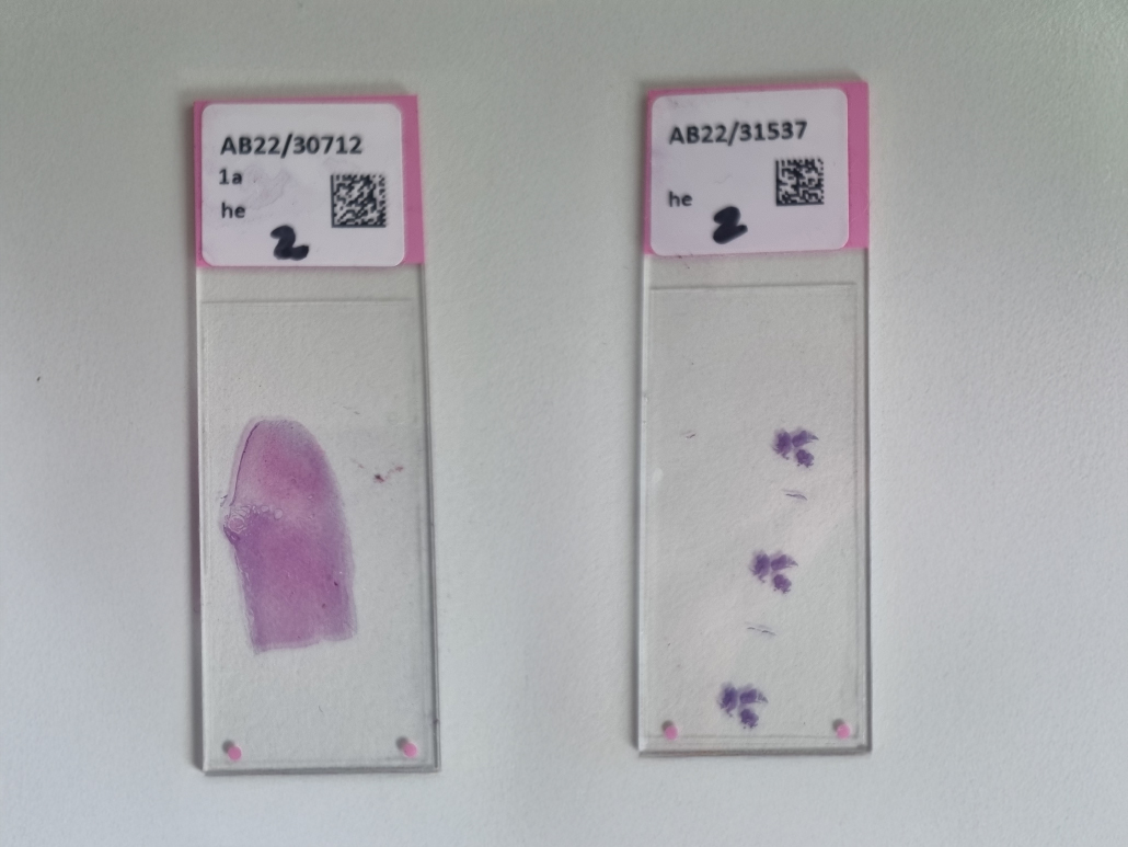
Figure 1. ‘Slide 1’ (left) and ‘Slide 2’ (right)
Manual Scanning
In addition to automatic scanners, there is also the option of manual scanning. These solutions are practical for quickly digitizing individual slides at low cost and sharing them, for example, via a cloud. Manual scan solutions are typically used in frozen section diagnostics or for tumor boards—anywhere where the purchase of an expensive scanner is not worthwhile, or where an automatic scanner might be slower. Manual scan systems can also be suitable for teaching/training, as the slide collection for education typically only needs to be scanned once.
While the main disadvantage is that these solutions are not suitable for scanning many slides, manual scan solutions offer some advantages over automatic scanners:
- Scanning with multiple objectives (automatic scanners typically only have one objective)
- Combining different magnification or staining levels on a single slide
- Low-maintenance system
- Scanning with immersion oil is possible
- Scanning with polarizer is possible
All manual programs work with a C-mount camera mounted on the microscope. A strong light source is necessary, preferably an LED, to achieve a constant color temperature. Additionally, a modern PC is required, as the software demands a solid CPU performance and memory (typically an i7 processor with 8–16 GB RAM).
All scan programs require the use of the mechanical stage, as they would not function properly due to potential rotational movements otherwise.
All programs operate on the same principle: By manually moving the slide, e.g., in a ‘meandering’ pattern, the camera captures multiple individual images and then stitches them together into a large image. This same principle is used in panorama shots taken with smartphones. Limitations of manual scanning include high magnifications (at 40x objectives, only partial scans are possible) and a large number of slides to be scanned.
However, scanning with oil, such as with a 40x high aperture objective (Zeiss), works brilliantly for hematology.
PathoZoom® Scan & LiveView
The Cologne-based company Smart In Media has developed a flexible solution that allows users to a) manually scan slides at their own microscope quickly (PathoZoom® Scan) and b) stream the microscope image over the internet (PathoZoom® LiveView). Additionally, Artificial Intelligence is already integrated and can be used in the form of ‘augmented reality’.
Here, we will focus only on PathoZoom® Scan.
The system is delivered in an attractive box—camera, software, user manual, and calibration slide are all included. After installing the camera and software, with the help of manuals, videos, and customer support, users can start scanning right away. PathoZoom® Scan impressed us with the highest scanning speed in our test, without generating artifacts. If the software loses track due to scanning too quickly, it can reconnect to the previously scanned area immediately once it’s found again.
One great feature: If an area was poorly focused (i.e., scanned out of focus), the same area can be revisited and refocused, with the software replacing the blurry section with the newly focused image
A ‘Freeze’ function allows users to pause scanning, move the current image, and then scan a different slide. This can be very useful, for example, to place an immunohistochemistry (IHC) slide next to a hematoxylin and eosin (HE) slide.
After scanning, the slide can either be saved locally in 3 open file formats (DICOM, TIFF, and SZI) or sent with a click through the integrated PathoZoom® SlideCloud via a secure link. The recipient doesn’t need an account or login and can immediately view the slide digitally via a browser. The annotation function even enables communication directly on the slide.
The software PathoZoom® LiveView included with the package is not discussed here, as it does not have scanning capabilities. It allows streaming of the microscope view over the internet and also offers Artificial Intelligence solutions.
Automatic Scanning
Anyone aiming to digitize a large number of slides (starting from 10 per day) or even fully digitize a pathology workstation (300 slides/day/pathologist) or an entire lab cannot avoid automation.
There are now many companies offering automated microscope scanners. However, the biggest obstacle to the widespread digitization of pathology remains the cost of these scanners. The author believes that if high-performance scanners were priced at around $50,000 USD, (almost) all pathology departments would move towards digitization.
A Word on Optics
Scanner manufacturers advertise their products with “20x” or “40x” scans. However, due to the different physical properties between analog microscopes and digital technology, differences arise that often cause confusion. When scanner manufacturers claim their scanner can scan at “40x,” they mean that the image impression corresponds to what a pathologist would see with a 40x objective lens and a 10x eyepiece—a total magnification of 400x. This is correct. However, the next step depends on the screen size and resolution.
Most scanners have a 20x objective lens installed to scan at 40x. This may seem counterintuitive at first, but it works because the sensor of the internal digital camera has a higher resolution than the human retina (in addition to other optical elements in the scanner and a very high numerical aperture).
Thus, magnification levels between scanners cannot be compared based on the objective lens alone, but only by looking at the microns per pixel (µm/px). Typically, 40x (400x magnification) corresponds to approximately 0.25 µm/px, 20x to 0.5 µm/px, 10x to 1.0 µm/px, and so on.
Test Results
For Occasional/Low-Volume Scanning
- Grundium Ocus
- Motic EasyScan One
- PathoZoom Scan & LiveView
For High-Throughput Scanning
- Hamamatsu S360 / Hamamatsu S20
- Leica Aperio GT450
- 3DHistech P1000 / P480
- KfBio 400
- Philips
- Argos
In terms of clinical routine experience, the following laboratories and scanners were available for testing:

| Scanner | Pathology Institute | Number of Scanners |
|---|---|---|
| Hamamatsu S360 | 6 | 10 |
| Hamamatsu S210 | 3 | 3 |
| Hamamatsu S20 | 2 | 4 |
| 3DHistech P1000 | 2 | 8 |
| Leica GT450 | 2 | 5 |
| Philips (old models, therefore excluded here) | 2 | 5 |
| Motic | 1 | 1 |
| KfBio | 1 | 1 |
| Argos 6x | 1 | 1 |
Small Scanners for Occasional Use
Grundium Ocus
The Finnish hardware manufacturer Grundium, founded by former Nokia employees, first impressed in 2018 with its compact and portable scanner, ‘Ocus’.
This is a 20x scanner (now also available in a 40x version), delivered in a practical carrying case. It can be quickly connected via Wi-Fi to any PC or tablet, which can then start the scan software through a web browser.
The scanner has a coarse focus adjustment, while fine adjustments are handled electronically via a built-in liquid lens.
The scan quality is excellent and comparable to larger devices. However, the scan time is significantly longer. For standard samples in our tests, the scan times were: 2:13 minutes (Slide 1) 6:32 minutes (Slide 2) for 40x scanning.
The main advantage of the scanner is its compact size, making it ideal for a personal desk scanner that delivers high-quality scans in various open file formats. It also supports integration with multiple cloud storage providers. Additionally, the live function allows the scanner to be remotely controlled. This means a technician can load a sample and adjust the focus at one location, while a pathologist views and analyzes it remotely. However, live microscopy is not equivalent to using a traditional microscope, as it takes some time for the scanner to move to the desired area, and only one magnification level can be used.
In our test lab, the initial enthusiasm for the Grundium Ocus was followed by a return to a manual Scan & LiveView solution for frozen section analysis, as the manual solution was simply faster in such situations (although not as high in resolution).
Nevertheless, the Grundium Ocus is an excellent piece of technology, well-suited for professional use. The price is approximately $18,000 (without taxes).
Motic EasyScan One
The Motic scanner is a compact device, available as a single-slide scanner or a six-slide version, with the smaller model being desk-friendly.
The scan quality is satisfactory, though it cannot compete with larger scanners from Hamamatsu, Leica, or 3DHistech. Additionally, the scanning speed is significantly slower.
For fast routine use, the device is only partially suitable, as slides must be placed in clamp holders, which is cumbersome. During our tests, some slides broke due to the size variance of slides in Germany (about 2 mm), which the clamp holders could not accommodate.
However, for those looking to digitize slides for teaching or research, the affordable price and acceptable quality make it a reasonable choice.
One drawback, common to many scanners, is its proprietary MDS image format, which is not widely recognized. However, it can be converted to other formats.
Professional Routine Scanners
In Germany and Europe, three main scanner companies dominate the market: Hamamatsu, 3DHistech, and Leica Aperio. All three produce high-quality scanners suitable for routine use, delivering excellent image quality and speed.
When asking pathologists and medical technical assistants, opinions are divided. Preferences for a particular device often come down to minor handling factors.
In general, when switching to digital scanning, an institute may need to adjust some workflow steps.
Hamamatsu NanoZoomer S360 & S20
The S360 is the largest scanner from the Japanese company Hamamatsu and the successor to the S210. The model number indicates its capacity: it can load 360 slides simultaneously. In contrast, the S20 has a loading capacity of only 20 slides, but these can be loaded directly from the Sakura staining rack. With the S360, slides must first be transferred into a proprietary Hamamatsu rack, adding extra time.
There is no difference in scan quality or speed between the models since the loading and scanning automation is the same, according to the manufacturer. The Hamamatsu S360 is widely used in Germany, and our experience with it has been extensive. The scan quality is excellent, and scanning speed is very high. Our tests showed impressive speeds of 0:41 min (Slide 1) and 1:07 min (Slide 2). Notably, there is no difference in speed between scanning at 20x or 40x magnification, though 40x results in larger file sizes.
The handling of the scanner is not as fully automated as, for example, the Leica GT450, since Hamamatsu requires a PC with its own scanner software for operation. However, compared to its predecessor, the S210, the S360 now features a well-functioning carousel loading system.
An additional traffic light indicator can be mounted on the scanner to alert technicians to problems. Over four years of daily use, only one major issue was observed: in laboratories with high dust levels, the scanner sometimes mistakenly identifies dust as tissue, significantly slowing down scanning as it attempts to scan empty areas. Regular dust cleaning is necessary to prevent this.
Another drawback is that the S360 cannot be loaded directly with racks from the staining bath, requiring an additional step of transferring slides into the Hamamatsu system. While this process is quick, it remains an extra step. A future model update is expected to address this.
With the S20, Hamamatsu introduced an innovative solution: most routine scanners process slides one at a time under a single objective lens. To speed up workflows, parallel scanning is required. This can be achieved by purchasing multiple S20 scanners, which are relatively small and more affordable than competitors, allowing multiple slides to be scanned simultaneously.
Leica GT450
The Leica Aperio GT450 is the flagship scanner from German company Leica, which acquired the American scanner manufacturer Aperio in 2012 to enhance and replace its own scanner series.
The Leica stands out with a modern design and compact size, making it suitable for placement on a standard lab bench. It features a built-in touchscreen for easy operation, eliminating the need for an extra PC with Windows applications for routine use. The scanner can be loaded directly from the Sakura staining bath using the same racks. Its carousel allows continuous loading, which is highly practical. The total capacity is 450 slides. The scan quality is good, though the scan time is slightly slower than the Hamamatsu S360/S20 and 3DHistech P1000.
During testing, the Leica Aperio GT450 proved to be a robust device for routine use. In a Hamburg laboratory using a Leica scanner alongside two 3DHistech P1000 units, preferences among technicians varied—some preferred the GT450, while others favored the P1000.
The only current drawback is the SVS file format. While implemented as an open standard in many systems, it is outdated since it only stores every second zoom level in image files, slowing down performance for web-based microscopy. The PathoZoom® Digital Lab diagnostic platform from Smart In Media optimizes SVS files into a faster open format. However, Leica recently also added support for exporting scans in the DICOM format.
Overall, this is a state-of-the-art system.
3DHistech Pannoramic P1000 and Pannoramic P480
The P1000 scanner is manufactured by Hungarian company 3DHistech and distributed in Germany by Sysmex and Epredia. It is by far the largest scanner among all manufacturers, requiring a space of 154 x 100 cm and weighing approximately 270 kg, making dedicated lab space necessary.
As the name suggests, the P1000 can load 1000 slides at once. However, in routine digitization, this capacity is less relevant since new slides are constantly being processed. Since unattended scans occasionally fail, it is questionable whether the full capacity will ever be fully utilized.
The scanner offers outstanding scan quality and recently performed best in a scanning test of blood and bone marrow smears without immersion oil, compared to the Leica GT450, Hamamatsu S360, and Motic EasyScan. A separate hematology test is available. 3DHistech is the only manufacturer to explicitly include cytology in its intended use documentation. It is the only system approved for cytology scans.
The system is easy to operate and can be loaded directly from the staining bath with Sakura racks, eliminating the need for slide transfers.
Scan speed is among the highest in the market, surpassing many competitors.
A smaller version, the P480, has a capacity of 480 slides but offers the same scan quality and speed.
A downside is the MRXS file format, which consists of an index file and around 20 additional files per slide. This makes file transfers cumbersome, particularly for uploading to online systems, as it is unclear when a transfer is fully complete. The SDK also had technical weaknesses, though 3DHistech has improved this by enabling scanning in the DICOM format. However, the DICOM format currently misses a directory table for the tiles, which lowers performance.
Philips IntelliSite
Philips scanners were the first microscope scanners approved by the FDA at an early stage for routine use. In this test, we had examined an initial model. Since this model has now become outdated and we did not find any pathology institute using one of the newer devices, we decided to forgo testing in this case.
It must be noted that Philips is the only scanner company mentioned here that offers a closed system. This means that the scanners cannot be purchased as standalone devices but must always be acquired together with the associated Image Management System and storage. Users have limited access to the digital slides without the Philips Image Management System, which in turn can hinder research projects and collaborations with other institutes. Many pathology departments have recognized this as a disadvantage, as they often prefer a combination of different scanners and image management systems.
KfBio 400
The Chinese company KfBio offers a full range of scanners, much like its Western competitors. Due to its unbeatable price of approximately $75,000 (net), we decided to test the KfBio 400, which has a capacity of 400 slides, in a pathology institute of one of our clients to evaluate whether this scanner could be added to our product portfolio alongside other offerings in 2022.
The initial enthusiasm upon unpacking the sleek, modern-looking system with a display and scanning software on a PC quickly gave way to disappointment. The English manual was not fully comprehensible, and it took several hours to get the scanner up and running with the help of online support. This was attributed to the lack of a European branch at the time of testing, which we initially noted as an area for improvement.
Disappointment turned into outright dismay when the lab’s IT department performed a virus scan on the included PC before integrating it into the laboratory network. The virus scan detected a Trojan horse hidden within a PDF document. As a result, we operated the scanner and PC only in a local installation within an isolated network.
The scanner itself delivers good image quality and demonstrated high scanning speed in this test. However, it repeatedly failed to detect loaded slides, an issue that even the technical online support was unable to resolve through software updates. Additionally, slides were frequently found dislodged inside the device. Moreover, the door can be opened while the carousel is rotating, which is a safety issue that, for example, is prevented in the Hamamatsu S360.
As much as we would have liked to find a cost-effective scanner for routine use to accelerate digitalization, this scanner was ultimately not viable for our pathologist and remained far from meeting European safety standards. However, as this dates back some time, we cannot do not know, if these issues have been solved.
Argos 6x
Behind the completely unique scanner Argos 6x lies a remarkable story. The German entrepreneur Thomas Gebing refused to accept that full automation in microscopic slide scanning was impossible simply because slides had to be manually loaded and unloaded. His goal was to acquire a digitalization technology to relieve his wife, dermatopathologist Dr. Ana Gebing, of some of her workload.
To achieve this, he founded the company Argos and initially developed an automation system using a Kuka robotic arm integrated into a large housing along with five single-slide scanners from another manufacturer. When their performance proved unsatisfactory, Argos promptly designed its own scanners. Six of these are now housed within the system, scanning slides in parallel while being loaded and unloaded by the robotic arm.
Several intelligent innovations were introduced in the process: racks can be loaded directly from the staining bath by simply placing them on a rail, allowing them to slide into position in front of the robotic arm via gravity. There is also an express track—a kind of secondary waiting position for urgent cases. Additionally, an intermediate archive is included: the robot first removes the scanned slides and places them on a shelf inside the housing. Only after a predefined waiting period do the slides move to their final destination—a box that can be easily swapped out. This design is both space-efficient and cost-effective.
A monitor mounted on the housing displays the scanning process of all six scanners simultaneously.
According to the manufacturer, the six scanners can scan 3,000 slides per day in parallel, or two slides per minute. However, the scanning speed of the individual scanners still has room for improvement, as the combined performance of all six is roughly equivalent to just two high-performance scanners from other manufacturers. That said, those other scanners do not offer full automation.
The resolution of the scanners has not yet reached 40x. In the slides we scanned, we found a resolution of 0.38 µm/pixel, which corresponds roughly to a 20x objective.
Anyone considering purchasing an Argos should be prepared to allocate sufficient space, as the system measures approximately 166 cm in width, 77 cm in depth, and 161 cm in height, with a weight exceeding 600 kg.
Scan Time Results:

| 3DHistech P1000 40x | 3DHistech P1000 80x | Grundium Ocus 40 40x | Hamamatsu S20/S360 40x | Leica GT450 | Motic 40x | PathoZoom® Scan | Ventana DP200 40x | |
|---|---|---|---|---|---|---|---|---|
| Slide 1 | 01:39 | 03:37 | 06:32 | 01:07 | 01:48 | 15:00 | 07:00 | 03:22 |
| Slide 2 | 00:36 | 00:53 | 02:13 | 00:41 | 01:46 | 12:00 | 09:00 | 02:34 |
Size and Weight Comparison of Scanners:

| 3DHistech P480 | 3DHistech P1000 | Grundium Ocus 40 | Hamamatsu S360 | Hamamatsu S20 | Leica GT450 | KfBio 400 | Motic EasyScan One | Argos 6x | |
|---|---|---|---|---|---|---|---|---|---|
| µm / pixel | 0,24 | 0,25 | 0,25 | 0,23 | 0,23 | 0,26 | 0,125 | 0,26 | 0,38 |
| Size (WxDxH in cm) | 120 x 90,2 x 100 | 154 x 98 x 90 | 18 x 18 x 19 | 75 x 69 x 62,8 | 49 x 62 x 45 | 52,8 x 71,1 x 49,5 | 52,4 x 71,1 x 49,5 | 20,5 x 39,8 x 42 | 116 x 77 x 161 |
| Weight (in kg) | 240 | 200 | 3,5 | 117 | 52 | 63,5 | 80,8 | 12,6 | 629 |
Conclusion
Pathologists looking to digitize their workflow for routine use will be well served by a scanner from Leica, Hamamatsu, or 3DHistech. These brands differ in price, ease of use, image formats, and support. However, none of them is significantly superior in speed or image quality to justify declaring a clear test winner. Hamamatsu and 3DHistech scanners are slightly faster than Leica’s, the latter offering a more user friendly and compact built. On a very positive note: these manufacturers are open to integrating with image management systems from other providers, which is a key requirement for successful digitalization.
Given the high cost of these devices, it is strongly recommended to observe them in real-time operation at a successfully digitized pathology lab and discuss their strengths and weaknesses with colleagues.
Regarding image formats, there is an encouraging trend toward the open medical DICOM standard, which will improve interoperability.
Scan Result Comparison
Now it’s your turn! Decide for yourself which scan results you find most convincing and compare the different scanners.